University of Texas at Austin

News

Willerson Center Postdoctoral Researcher Wins NIH Fellowship

By Rebecca Riley

Published Oct. 10, 2022

An aortic cell illustration being stretched. Credit: Toni West and Don Howsmon.

Toni West, currently a postdoctoral research fellow at the Oden Institute's Willerson Center for Cardiovascular Modelling and Simulation, has been awarded the Ruth L. Kirschstein postdoctoral fellowship from the National Institutes of Health

This opportunity is designed to support highly promising postdoctoral candidates with the potential to significantly advance the study of health. Dr. West was chosen for her research on uncovering the cellular basis for aortic valve disease. In receiving her fellowship, she became the third NIH postdoctoral fellow in the Willerson Center, alongside Drs. Christian Goodbrake and Dan Howsmon.

The Willerson Center is so named in honor of late American cardiologist, James T Willerson, who was the President Emeritus, Director of Cardiology Research, and Co-Director of the Cullen Cardiovascular Research Laboratories at the Texas Heart Institute. 

“These are very competitive awards and speak volumes about Dr. Willerson’s vision, the Oden Institute, and the greater research environment at The University of Texas at Austin, ” said Director of the Willerson Center, Michael Sacks.

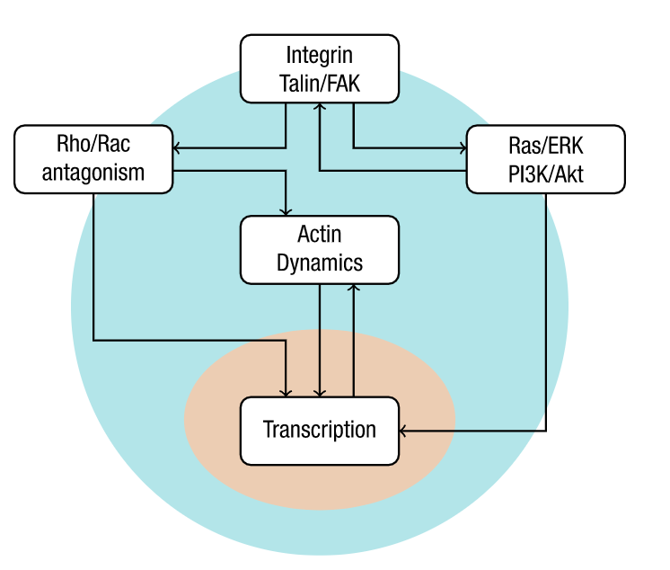
Every time a human heart beats, its aortic valves open and close – over 3 billion times in a typical lifetime. Though impressively durable, aortic valves can become diseased and require surgical replacement – a procedure that continues to have problems with regards to long-term durability.
 

These are very competitive awards and it speak volumes about Dr. Willerson’s vision, the Oden Institute, and the greater research environment at UT Austin.

— Michael Sacks, Director of the Willerson Center.

To allow for the development of more permanent non-surgical treatments, Drs. Toni West and Dan Howsmon are creating the first computational models of the complex signaling interactions that occur in cells in the aortic valve in an effort to explore the relationship between them and aortic valve disease.

“The dynamic forces that occur in the aortic valve are enormously important for maintaining a normal aortic heart valve,” said West. “Yet, most of the research to develop pharmacological therapies for aortic valve disease has used cells lying still in a petri dish.”

Using real clinical images, Willerson researchers have developed computational techniques that have uncovered specific stretch patterns associated with the motion of healthy and diseased aortic valves. With the help of Willerson Center affiliated faculty, Dr. Aaron Baker– based in the Department of Biomedical Engineering at UT Austin’s Cockrell School of Engineering  - Toni West asks what it is about these stretch patterns that correlate with healthy aortic heart valve cell function.

block.caption

Illustration of the key signaling modules involved in VIC mechanobiology. Credit: Oden Institute

“Is it the slope of a stretching waveform that makes the cells act the way they do, or is it the overall average amount of stretch that they get, or is it the peak rate that they get? Which part of the waveform is most important for whether the cell becomes activated into an unhealthy state? That is the question,” explained West.

To find the answer, she uses computer simulations to analyze the cells for effects of the stretching process. This information is then fed into Dr. Howsmon’s computational cell signaling models, identifying important pathways of how the cells function.

“The first step is to analyze how the cells have been affected,” said West. “The next step will be to figure out how biochemicals present in the diseased aortic heart valve affect these signaling patterns. In the long-term, we’ll be able to identify targets to find a drug to treat the disease itself.”

Treating aortic valve disease is not the only potential future impact of this research. Another is forecasting how the disease would respond to surgical intervention. With the images from 3D  echocardiography, computational models can be developed to make predictions on a patient-by-patient basis.

“With new machine learning methods, we can compute the forces inside the heart valve fast enough to inform decisions on the clinical level,” said West. “In all of this work, it’s really important to be collaborating with others on these big issues, working towards developing improved therapies that will allow patients to enjoy an improved quality of life.”